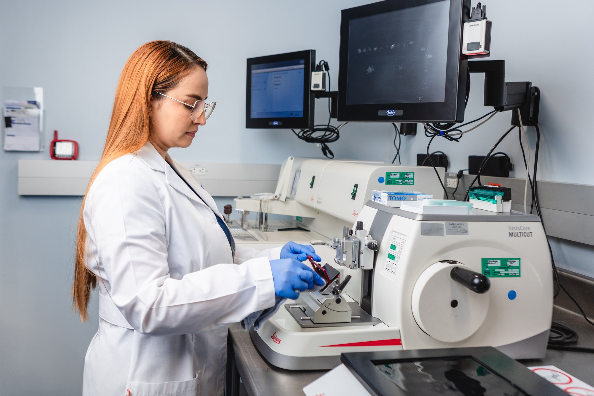
Pathology Laboratory
The laboratory specializes in accurate diagnosis of diseases affecting tissues and organs through biopsies and cytology.
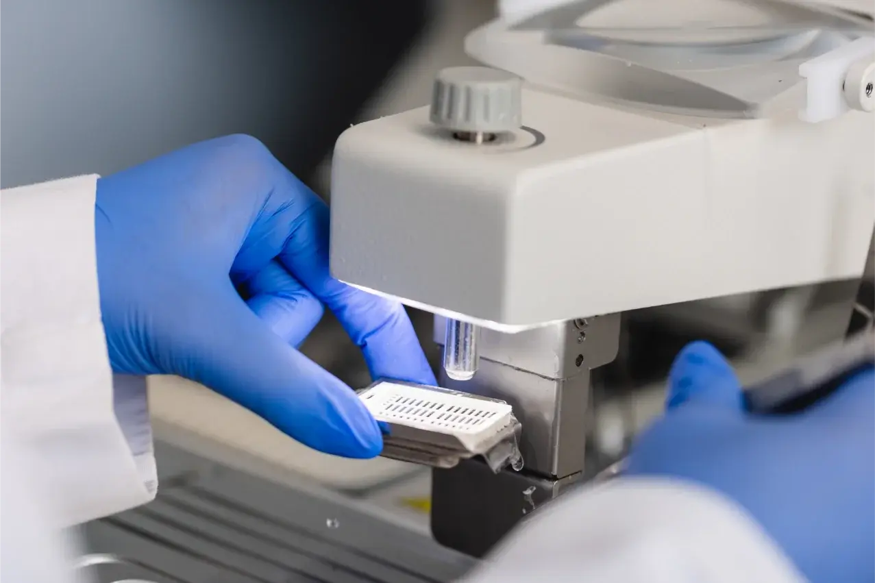
We have automated equipment to perform specialized studies to provide more accurate and complete diagnoses.
Procedures such as transoperative freezing, Mohs surgery and ROSE Rapid On-Site Evaluation.
We innovate in the detection of HPV-associated cervical cancer with advanced technology, including:
- Liquid-based cytology.
- p16 immunohistochemistry
- Dual staining
- Cobas 5800
Available in:
Santa Ana facility
Sede facility
Aleste facility
Hours of Operation
Monday to Friday from 7:00 a.m. to 5:00 p.m.
For procedures in the evenings, weekends and holidays, prior coordination with the laboratory is recommended.
In case of an emergency intraoperative consultation without prior coordination, attendance will depend on the availability of the pathologist and technician.
Biopsies
Study of tissues taken during medical procedures to identify abnormal conditions.
How is tissue processed in the pathology laboratory?
This process ensures that the quality of the sample is preserved and that accurate and reliable results are generated for timely and effective diagnosis.
Sample reception
The tissue or cell sample arrives at the laboratory properly labeled with patient information and site of origin.
Fixing
The sample is placed in formalin to preserve its structure and avoid decomposition.
Processing
The tissue goes through a series of solutions that prepare it to be embedded in kerosene. This gives the tissue the right consistency to be cut into extremely thin sections.
Cut
Using a microtome, the tissue is cut into thin layers, approximately 4 micrometers thick (thinner than a human hair).
Staining
The sections are stained with special stains (such as hematoxylin and eosin) that highlight the cellular structures, facilitating observation under the microscope.
Evaluation
The pathologist examines the stained specimens under a microscope, looking for abnormalities or specific features that indicate disease or pathologic changes.
Diagnostic report
Based on the microscopic observations and, in some cases, additional studies such as immunohistochemistry or molecular studies, the pathologist prepares a detailed report with the final diagnosis.
Cytologies
Evaluation of cells for possible changes associated with disease, such as cancer or infections
How is a liquid-based cytology processed in the pathology laboratory?
The analysis of a liquid-based cytology follows a specific process that optimizes the collection and analysis of cells, improving the quality of diagnosis and reducing the need for repeat testing.
Sample reception
Cells are collected from the area of interest (e.g., cervix for cervicovaginal cytology) using a brush, and then transferred to a bottle with a special liquid solution that preserves the cells.
Laboratory processing
the liquid sample is agitated to disperse the cells, removing impurities such as mucus, blood or unwanted cellular debris. This improves the quality of the sample for analysis.
Slide preparation
Through an automated process, a thin layer of cells is transferred from the liquid solution to a glass slide, creating a uniform and easy-to-evaluate sample.
Staining
The cell sample is stained with special dyes that allow differentiation of the different cell structures and detection of any abnormalities, such as precancerous or infectious changes.
Microscopic evaluation
The pathologist examines the specimen under a microscope, evaluating the characteristics of the cells for signs of infection, inflammation or precancerous changes that may require follow-up.
Diagnostic report
Based on the cytology observations, the pathologist prepares a detailed report that will be sent to the treating physician for decisions on appropriate clinical management.
Virtual Tour

Having a sample traceability system in the pathology laboratory is crucial to ensure safety and accuracy at every stage of the diagnostic process.
Automation offers multiple advantages, such as greater precision, speed and reproducibility of results. It allows a larger volume of samples to be processed with a smaller margin of human error, resulting in more reliable diagnoses and shorter times. In addition, it is essential that physicians and patients know the type of tests and equipment used, as this guarantees the transparency and quality of the diagnostic process, ensuring the use of advanced and validated technologies for the timely detection of diseases.
This technique allows us to identify specific proteins in tissues. It is a key tool for diagnosing different types of cancer and classifying tumor subtypes.
We have a complete panel for the diagnosis of breast cancer that includes the evaluation of estrogen receptors, progesterone receptors, the Ki-67 cell proliferation index and the HER2 protein.
In addition, we offer the HER2 Dual ISH DNA technique, a sophisticated in situ hybridization method, essential in the evaluation of HER2 gene status in breast cancer.
For antibody analysis in biopsies with suspected melanoma, we use chromogen red studies. This technique improves the detection of antibody expression, avoiding confusion with melanin deposition in the tumor.
We perform microsatellite instability studies in colon cancer cases. This evaluation is essential to determine microsatellite status and can help in the identification of patients who might benefit from specific treatments.
A method that allows the detection of genetic alterations directly in the tissue. It is used in the identification of certain genes associated with different types of cancer, such as breast cancer.
Used to identify the presence of antibodies or antigens in tissue by the application of fluorescent dyes. Useful in the diagnosis of autoimmune diseases or skin disorders.
This technique allows a rapid diagnosis to be obtained during surgery. The tissue is immediately frozen and examined under the microscope so that the attending physician can make immediate decisions during the procedure.
We evaluate samples in real time during needle puncture procedures. This guarantees correct sample collection, ensuring a more accurate diagnosis and reducing the need for repeat procedures.
We perform molecular analysis on samples previously processed in kerosene, which allows us to study genetic mutations and molecular alterations. This is key to design specific treatments for different types of cancer.
Real-time or digital PCR Cancer Tests- Epidermal growth factor receptor (EGFR)
- KRAS
- NRAS
- BRAF
- PIK3CA
- JAK2 (for hematology cases)
NGS Cancer Testing on Illumina platform
- BRCA 1 and 2
Studies of Microorganisms on Paraffin Tissue
- Tuberculous and atypical mycobacteria
- Aspergillus sp.
- HPV
- Pneumocystis jirovecii
- Toxoplasma gondii
We offer special stains to highlight specific cellular and tissue structures. Fundamental for the diagnosis of infections, fibrosis, and other specific pathologies.
|
Immunohistochemistry (IHC) |
|||||
|---|---|---|---|---|---|
|
AMACAR |
Mammaglobin |
P60 |
|||
|
SMA |
CDX2 |
Her2 Dual CISH DNA |
|||
|
AML |
CEA |
HMB45 (red chromogen) |
|||
|
Basal Cell |
CK 5/6 |
Ki 67 |
|||
|
BCL2 |
CK19 |
E-Cadherin |
|||
|
BCL6 |
CK20 |
GATA3 |
|||
|
Caldesmon |
CK7 |
PanCK |
|||
|
Calretinin |
Chromogranin |
PAX-5 |
|||
|
CD5 |
Cyclin D1 |
PAX-8 |
|||
|
CD10 |
Desmin |
PMS2 |
|||
|
CD117 |
DOG-1 |
Podoplanin (D2-40) |
|||
|
CD20 |
EMA |
PSA |
|||
|
CD23 |
Melan-A (red chromogen) |
Estrogen Receptors |
|||
|
CD3 |
MHS 2 |
Progesterone Receptors |
|||
|
CD30 |
MHS 6 |
S100 |
|||
|
CD31 |
MLH-1 |
Synaptophysin |
|||
|
CD34 |
MUM-1 |
SOX10 (option with red chromogen) |
|||
|
CD45/LCA |
P16 (E6H4) |
TTF-1 |
|||
|
CD5 |
P40 |
Vimentin |
|||
|
CD68 |
P53 |
WT1 |
|||
|
HER2/ NEU (4B5) |
GFAP |
||||
|
Immunofluorescence |
|
C1q |
|
C3 |
|
Fibrinogen |
|
IgA |
|
IgG |
|
IgM |
|
Kappa |
|
Lambda |
Pathology: Web Form
We want to assist you before, during and after your visit. Find everything you need to make your hospital stay as comfortable as possible.
We have a specialized insurance team at your disposal. If you have any questions about requirements or need support contact us.
All payments for outpatient, inpatient and surgical services must first be verified by means of the statement of account at the Department of Patient Management and Formalization and then be paid at the cashier's office in the same department.
Patient Information
Insurance
We make using your medical insurance simple. Your time is valuable, so we manage your authorizations efficiently.